Labeled Diagram Of An Eye - Diagram Of Eye Labelled Diagram Of Human Eye Explanation And Function / Anatomy of the eye labeled.
byAdmin-
0
Labeled Diagram Of An Eye - Diagram Of Eye Labelled Diagram Of Human Eye Explanation And Function / Anatomy of the eye labeled.. The anterior part of this layer is called cornea. Eye anatomy, function, and physiology facts. Anatomy of an eye diagram application note tektronix september 2010, but end up in harmful downloads. It is the transparent membrane which refracts the light entering our eye. A human eye is roughly 2.3 cm in diameter and is almost a spherical ball filled with some fluid.
Parts of the eye, eye diagram, vitreous gel, iris, cornea, pupil, lens, optic nerve, macula, retina created date: This is a small tube that runs from the eye to the nasal cavity. In case you need a little refresher before going over your lesson, or want something for your slightly older children to read, we have added a simply worded, but terminologically accurate summary, describing the. From the center of the optic nerve radiates. The anterior part of this layer is called cornea.
Structure And Function Of The Human Eye from www.thoughtco.com The eye sits in a protective bony socket called the orbit. For us to see, there has to be light. The human eye is composed of many different parts that work together to interpret the world around us. Label parts of the human eye. Lungs and respiratory system for kids gcse science breathing and. The eye is rather like a living camera. Parts of the eye outside the eyeball. Fungi diagram gcse top electrical wiring diagram.
Rather than enjoying a good pdf once a cup of coffee in the afternoon,
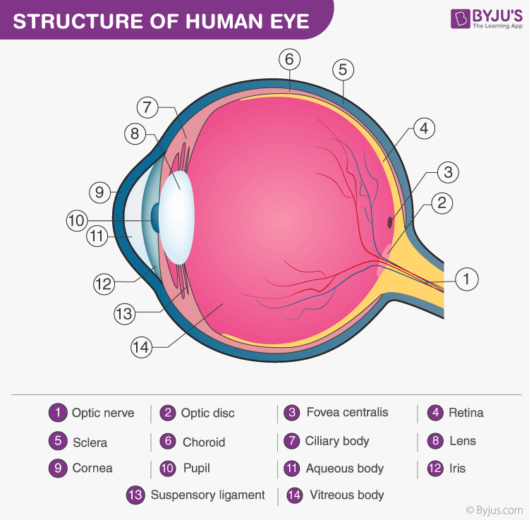
The extraocular muscles are attached to the white part of the eye called the sclera. For us to see, there has to be light. Human eye anatomy quiz diagram labeling, eye anatomy model, interactive eye diagram quiz. The cornea, the pupil, the iris, the lens, the vitreous humor, the retina, and the sclera. This is why a teary eye is usually accompanied by a runny nose. The front transparent part of the sclera is called cornea. Human eye consists of various parts which helps us in seeing the objects, the function of various parts are: Iris controls the size of pupil. An eye diagram triggered from a clock recovered from the data signal using a narrow loop bandwidth clock recovery scheme. Let's take a closer look at each of these. Contrary to popular belief, the eyes are not perfectly spherical; Our eyes are highly specialized organs that take in the light reflected off our surroundings and transform it into electrical impulses to send to the brain. A human eye is roughly 2.3 cm in diameter and is almost a spherical ball filled with some fluid.
You should make a label that represents your brand and creativity, at the same time. This is a small tube that runs from the eye to the nasal cavity. Often called lazy eye, this condition starts in childhood.one eye sees better than the. Iris controls the size of pupil. The eye diagram to label teaching resources.
Structure And Function Of The Human Eye from www.thoughtco.com Facts about the eye to understand more in detail about our eye and how our eye functions, we need to look into the structure of the human eye. For us to see, there has to be light. Diagram of the eye diagram of eyeball cross section of human eye section of human eye cross section eye cross section of eye anatomy of the eye eye parts human eye diagram of eye. This is why a teary eye is usually accompanied by a runny nose. The extraocular muscles are attached to the white part of the eye called the sclera. Anatomy of the eye labeled. Let's take a closer look at each of these. In case you need a little refresher before going over your lesson, or want something for your slightly older children to read, we have added a simply worded, but terminologically accurate summary, describing the.
From the center of the optic nerve radiates.
This helps us to understand how each one is situated and related to the other. Instead, it is made up of two separate segments fused together. The anterior part of this layer is called cornea. National eye institute , national eye health education program subject: At the front of the eye is a clear, round window called the cornea. One of our favorite ways to get to grips with all of the parts of the eye is by utilizing labeled diagrams. This is a small tube that runs from the eye to the nasal cavity. Our eyes are highly specialized organs that take in the light reflected off our surroundings and transform it into electrical impulses to send to the brain. The core eye anatomy diagram, designed as the labeling exercise, has a fully colored and labeled reference chart to go with it. It is the external layer composed of dense connective tissue. An easy and convenient way to make label is to generate some ideas first. The iris of the eye functions like the diaphragm of a camera controlling the amount of light reaching the back of the eye by automatically adjusting the size of the pupil aperture. The cornea, the pupil, the iris, the lens, the vitreous humor, the retina, and the sclera.
Causes loss of central vision as you get older. An incomplete eye diagram formed by triggering on data. With help from other important structures in the eye, like the iris and cornea, the appropriate amount of light is. Human eye consists of various parts which helps us in seeing the objects, the function of various parts are: The anatomy of the eye is fascinating, and this quiz game will help you memorize the 12 parts of the eye with ease.
Structure And Functions Of Human Eye With Labelled Diagram from cdn1.byjus.com This is why a teary eye is usually accompanied by a runny nose. The front transparent part of the sclera is called cornea. See human eye diagram stock video clips. With help from other important structures in the eye, like the iris and cornea, the appropriate amount of light is. Human eye anatomy quiz diagram labeling, eye anatomy model, interactive eye diagram quiz. Draw a well labelled diagram of eye. Diagram of human eye with labelling. Labeled diagram of the eye
Facts about the eye to understand more in detail about our eye and how our eye functions, we need to look into the structure of the human eye.
Lungs and respiratory system for kids gcse science breathing and. For us to see, there has to be light. Six extraocular muscles in the orbit are attached to the eye. The cornea, the pupil, the iris, the lens, the vitreous humor, the retina, and the sclera. From the center of the optic nerve radiates. See human eye diagram stock video clips. It is the external layer composed of dense connective tissue. Eye anatomy, function, and physiology facts. It is the transparent membrane which refracts the light entering our eye. This is a small tube that runs from the eye to the nasal cavity. Labeled diagram of the eye You should make a label that represents your brand and creativity, at the same time. Although the eye is small, only about 1 inch in diameter, each part plays an important role in allowing people to see the world.

/GettyImages-695204442-b9320f82932c49bcac765167b95f4af6.jpg)